Embryo with Yolk sac in First Trimester
From researchgate.net
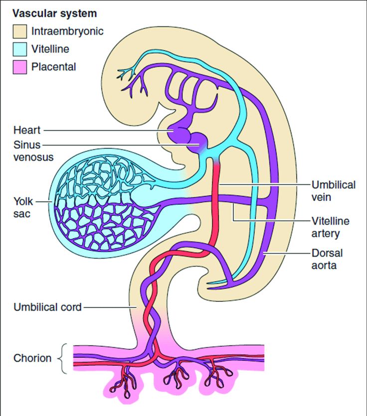
Diagram
- Vascular refers to "Blood vessels"
- Dorsal aorta "Back artery"
- Umbilical arteries (purple) vein (red)
- Vitelline a Latin word for "Egg yolk"
- Vitelline arteries are arteries that bring blood to the yolk sac
- Vitelline veins are veins that drain blood from the yolk sac
- Diameter c. 5mm
The vitelline and umbilical veins begin as pairs of symmetrical vessels that drain separately into the sinus venosus of the heart. Over time, these vessels become intimately associated with the rapidly growing liver.
Initially blood is conveyed to the wall of the yolk sac by the vitelline arteries (a branch of the dorsal aorta).
After circulating through a wide-meshed capillary plexus, it is returned by the vitelline veins to the tubular heart of the embryo.
This constitutes the vitelline circulation, and by means of it nutritive material is absorbed from the yolk-sac and conveyed to the embryo.
Much of it is incorporated into the primordial gut during the fourth week of development.
By 10 - 12th week placenta has taken over.
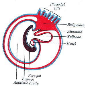
During week 3, a finger-like outpocketing of the yolk sac develops into the allantois, a primitive excretory duct of the embryo that will become part of the urinary bladder. Together, the stalks of the yolk sac and allantois establish the outer structure of the umbilical cord.
|
Later stage of allantoic development with commencing constriction of yolk-sac
Click image for full screen
|
|
Further notes on the umbilical cord
Extract from ncbi.nlm.nih.gov/books
- The development of the umbilical cord begins in the third week of embryologic formation. The developing embryo consists of a three layer disc - the ectoderm (outer layer), mesoderm (middle layer), and endoderm (inner layer) attached to the decidua basalis (the mucous membrane that lines the uterus) by the connecting stalk, the primitive umbilical cord. The connecting stalk is a thick stalk of the extraembryonic membrane extending from the caudal (tail) end of the embryo to the center of the developing placenta on the decidua basalis.
- The process of body folding occurs during week four with rapid growth amnion and embryonic disc compared to the yolk sac. Cranial caudal (head and tail) folding causes approximation of the connecting stalk and yolk sac on the ventral (belly) surface of the embryo. The amnion expands to cover the entire embryo except for the rudimentary umbilical ring, where the connecting stalk and yolk sac emerge. During this time, the allantois (from Greek word for sausage) an outpouching of the endodermal hindgut, forms and extends into the connecting stalk for the purpose of exchanging gases and handling liquid waste. It evolves into the urachus, a channel between the bladder and the umbilical cord that usually seals off and obliterates around the 12th week of gestation leaving a small fibrous cord between the bladder and belly button, the median umbilical ligament.
- Between the fourth and eighth weeks, there is an increase in amniotic fluid production, which causes the amniotic cavity to swell and fill the chorionic space. This increase in the amniotic fluid also causes elongation of the connecting stalk, and the yolk sac is compressed down within the connecting stalk to form the vitelline duct. The expansion of the amniotic cavity causes the amnion and the chorion to come into contact, and the extraembryonic mesoderm covering these two layers fuses. As such, the chorionic cavity disappears, leaving the umbilical cord surrounded by the amnion, floating in the amniotic fluid.
- By week seven, the intestines begin to herniate out of the embryo through the umbilical ring and into the umbilical cord. This physiologic herniation is necessary for proper rotation of the intestines and adequate growth of the fetus to house the expanding intestines. The rapid development of the intestines causes elongation of the umbilical cord.
- Between weeks ten and twelve, the intestines leave the umbilical cord and return to the abdominal cavity. The umbilical cord continues to elongate during the second trimester with a length comparable to the crown-rump length of the fetus.
- By term, the vitelline duct and allantois have typically completely involuted. However, in some cases, remnants of the allantois and vitelline duct can be found in the umbilical cord proximal to the neonate. At birth, the cord typically measures an average of 50 to 60 cm in length and 2 cm in diameter with up to 40 helical turns. After the birth of the neonate, the umbilical cord is clamped and then cut as the neonate now breathes on its own, and the remainder of the umbilical cord is delivered along with the placenta.
** End of page