Baby first few weeks in the Womb
Extract from courses.lumenlearning.com /boundless-ap/chapter/ first-week-of-development/
Following fertilization a series of rapid cell divisions occur that decreases each cell’s size with each subsequent division — this eventually produces a morula. The different cells derived from cleavage, up to the blastula stage, are called blastomeres.
The morula in humans is a solid ball of about 16 undifferentiated, spherical cells. The morula enters the uterus about four days after fertilization and "floats" in the uterine cavity. During this time, it continues to divide and absorbs fluid from the uterine cavity. With this cell division, the blastomeres change their shape and tightly align themselves against each other. This is called compaction.
As the fluid collects between the trophoblast (the outer or peripheral layer which later forms the placenta) and the greater part of the inner cell mass (which subsequently forms the embryo), the morula is converted into the blastocyst (or blastula).
In humans, the blastocyst is formed approximately five days after fertilization.
The end of cleavage, at over 100 cells is known as the midblastula transition and coincides with the onset of zygotic transcription, at which development comes under the exclusive control of the zygotic genome rather than the maternal (mum) genome.
Second week
In humans, implantation of a fertilized ovum occurs between 6 to 12 days after ovulation.
In preparation for implantation, the blastocyst sheds its outside layer, the zona pellucida (shell), and is replaced by a layer of underlying cells called the trophoblast. The trophoblast will give rise to the placenta after implantation.
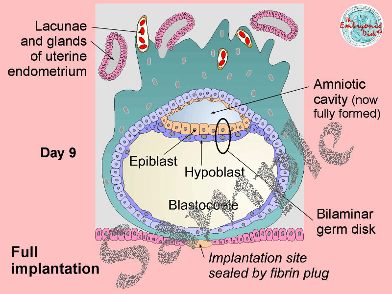
The inner cell mass is composed of two kinds of cells:
- the epiblast that forms the embryo, referred to as embryonic stem cells (ESCs)
- the hypoblast that will form the chorion i.e. the outermost fetal membrane, the amniotic membranes, and also the cells of the primary yolk sac, a membranous sac attached to the embryo for nutrition. Also called the umbilical vesicle
See an image below of the embryo at Day 9 from ucl.ac.uk with the "blastocoele" cavity, which will become the yolk sac.
 Third week
During the third week, as the heart is built, the yolk sac forms primitive blood islands.
Fourth week
During the fourth week much of the primary yolk sac is "pinched off" and incorporated into the primordial gut. It is now known as the secondary yolk sac. The embryo is about 4 mm.
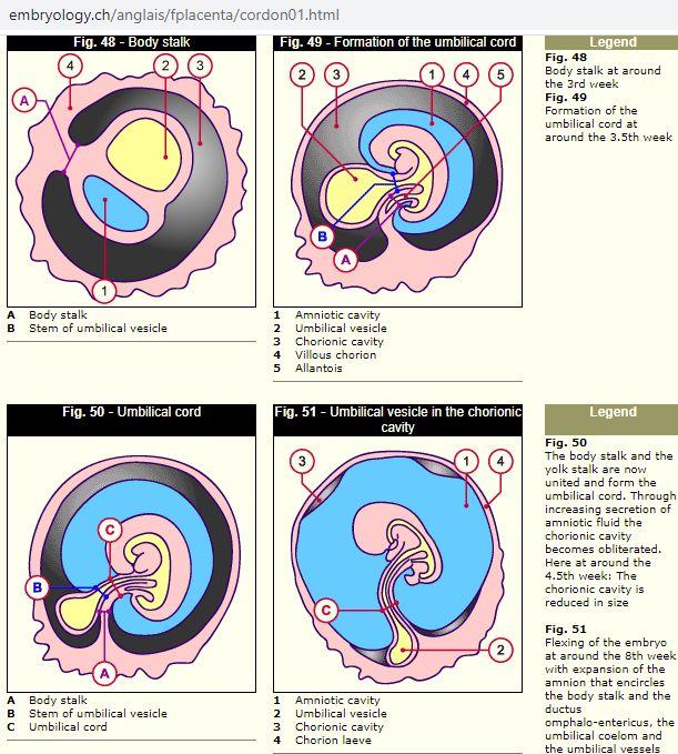
See the image below of Week 3, Week 3½, Week 4½, and Week 8 when the embryo is about 2 cm. The yolk sac is coloured yellow, the amniotic cavity is coloured blue.
Note too that in the first image (in Week 3) the cells that will form the tail of the embryo are at the top and the head is at the base.
Third week
During the third week, as the heart is built, the yolk sac forms primitive blood islands.
Fourth week
During the fourth week much of the primary yolk sac is "pinched off" and incorporated into the primordial gut. It is now known as the secondary yolk sac. The embryo is about 4 mm.
See the image below of Week 3, Week 3½, Week 4½, and Week 8 when the embryo is about 2 cm. The yolk sac is coloured yellow, the amniotic cavity is coloured blue.
Note too that in the first image (in Week 3) the cells that will form the tail of the embryo are at the top and the head is at the base.
 Blood formation in the liver begins about the 5th week, then a regular heartbeat by the 6th week. In the 12th week blood formation will also occur in the spleen and lymph nodes, then when bone marrow develops in the second trimester, it takes over much of the task.
About the 10th week when the embryo is about 8 cm, the mother's placenta takes over nutrition.
Click here for further notes.
Amniotic Fluid starts as water and electrolytes (Na+ K+ Ca+ Cl-), about 25ml, generated from maternal plasma through the fetal membranes.
By about the 12-14th week the liquid also contains proteins, carbohydrates, lipids and phospholipids, and urea. About 10% - 14% of its nutritional requirements.
From week 16, includes fetal urine.
At week 20, about 400ml
At birth, 800ml - 1000ml.
Blood formation in the liver begins about the 5th week, then a regular heartbeat by the 6th week. In the 12th week blood formation will also occur in the spleen and lymph nodes, then when bone marrow develops in the second trimester, it takes over much of the task.
About the 10th week when the embryo is about 8 cm, the mother's placenta takes over nutrition.
Click here for further notes.
Amniotic Fluid starts as water and electrolytes (Na+ K+ Ca+ Cl-), about 25ml, generated from maternal plasma through the fetal membranes.
By about the 12-14th week the liquid also contains proteins, carbohydrates, lipids and phospholipids, and urea. About 10% - 14% of its nutritional requirements.
From week 16, includes fetal urine.
At week 20, about 400ml
At birth, 800ml - 1000ml.
** End of page